Currently Empty: £0.00
mbakr
- November 21, 2025
- Comment 0
ABSTRACT
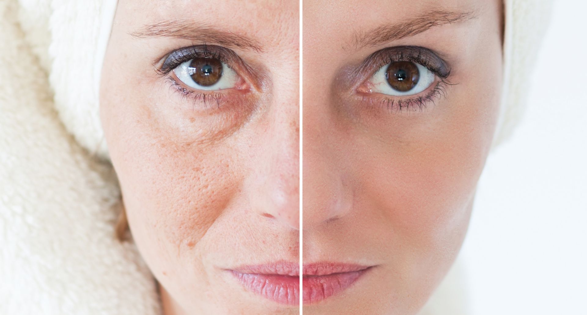
Hyperpigmentation is one of the most common skin concerns worldwide, with causes ranging from UV exposure and inflammation to hormonal imbalance and genetic predisposition. Its management requires a deep understanding of melanocyte biology, pigment pathways, clinical presentation, and treatment strategies tailored to diverse skin phototypes. This module provides an in-depth overview of the pathophysiology of hyperpigmentation, diagnostic approaches, classification of pigmentary disorders, and evidence-based treatment algorithms including topical agents, chemical peels, lasers, light devices, and regenerative therapies. Designed for aesthetic and dermatology practitioners, it emphasizes safe, structured, and effective management of hyperpigmentation across global patient populations.
LEARNING OBJECTIVES
Upon completing this module, learners will be able to:
- Understand melanocyte biology and mechanisms of pigment production.
- Identify and classify common pigmentary disorders.
- Apply diagnostic tools such as Wood’s lamp and dermatoscopy.
- Develop individualized treatment plans based on skin type and pigmentation depth.
- Implement evidence-based therapies including topicals, peels, lasers, and combination regimens.
- Prevent and manage complications such as PIH and rebound pigmentation.
- INTRODUCTION
Hyperpigmentation affects all skin types but is especially prevalent in medium to dark phototypes (Fitzpatrick IV–VI). It can significantly impact quality of life and is one of the leading reasons patients seek dermatologic and aesthetic care.
Common forms include:
- Melasma
- Post-inflammatory hyperpigmentation (PIH)
- Lentigines
- Freckles
- Dermal melanocytosis
- Drug-induced pigmentation
Successful treatment requires identifying the type, depth, and triggers of pigmentation and tailoring therapy to the patient’s skin phototype.
- MELANOCYTE BIOLOGY & PATHOPHYSIOLOGY
2.1 Melanin Production
Melanin is produced by melanocytes in the basal epidermal layer.
Two types:
- Eumelanin (brown/black; protective)
- Pheomelanin (yellow/red; less protective)
2.2 Melanogenesis Process
- Tyrosine → DOPA (via tyrosinase)
- DOPA → Dopaquinone
- Final melanin polymer formation
Tyrosinase is the rate-limiting enzyme, targeted by most pigment-control therapies.
2.3 Causes of Pigment Overproduction
- UV Exposure
- DNA damage triggers melanogenesis
- Increases tyrosinase activity
- Causes lentigines and diffuse darkening
- Inflammation
- Post-acne
- Post-eczema
- Post-procedural (lasers, peels)
→ Leads to PIH
- Hormonal Factors
- Pregnancy
- Birth control
- Thyroid imbalance
→ Major driver of melasma
- Genetics
Freckles and some dermal pigmentation types.
- Medications
- Antimalarials
- Chemotherapy agents
- Hormones
- CLASSIFICATION OF HYPERPIGMENTATION
3.1 Epidermal
- Melanin in the basal/suprabasal layers
- Appears brown
- More responsive to treatment
3.2 Dermal
- Melanin-laden macrophages deeper in dermis
- Appears blue-gray
- Harder to treat
3.3 Mixed
Most melasma cases.
- COMMON PIGMENTARY DISORDERS
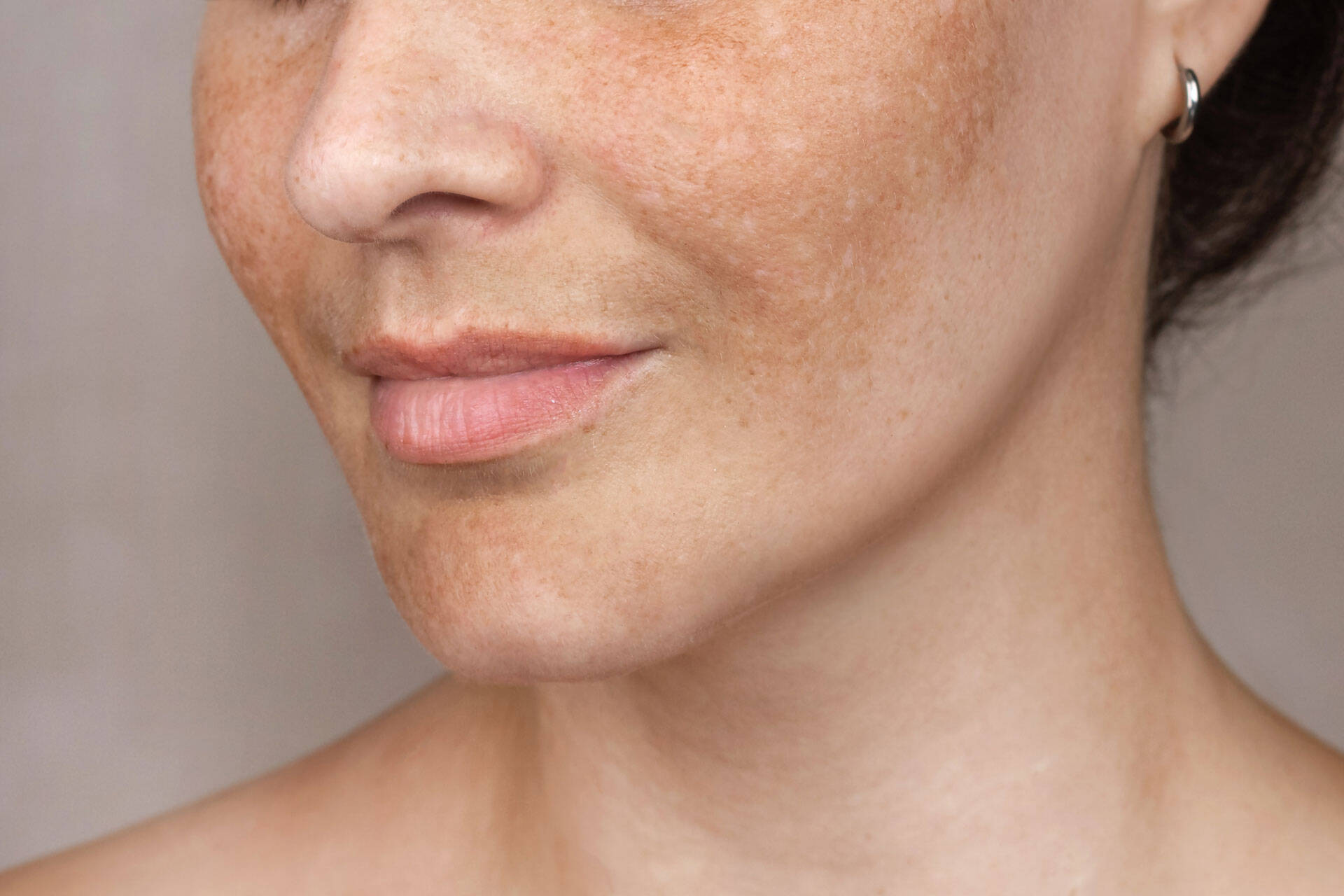
4.1 Melasma
Chronic, relapsing hyperpigmentation.
Common patterns:
- Centrofacial
- Malar
- Mandibular
Triggers:
- Hormones
- UV exposure
- Heat
- Inflammation
4.2 Post-Inflammatory Hyperpigmentation (PIH)
Darkening following:
- Acne
- Eczema
- Injuries
- Procedures
Common in Fitzpatrick IV–VI.
4.3 Lentigines
Solar-induced lesions.
Characteristics:
- Sharply demarcated
- Result of chronic UV damage
4.4 Freckles (Ephelides)
Genetic tendency.
Darken with sun exposure.
4.5 Drug-Induced Hyperpigmentation
From:
- Minocycline
- Amiodarone
- Antimalarials
- Cytotoxic drugs
4.6 Dermal Melanocytosis
Examples:
- Hori’s nevus
- Nevus of Ota
Requires specific lasers.
- DIAGNOSTIC APPROACH
5.1 Patient History
Assess:
- Onset
- Triggers
- Sun exposure habits
- Hormonal history
- Product use
- Previous procedures
5.2 Skin Examination
Look for:
- Color
- Borders
- Distribution
- Symmetry
5.3 Wood’s Lamp
Helps determine depth.
- Epidermal → enhances
- Dermal → no enhancement
5.4 Dermatoscopy
Useful for:
- Melasma patterns
- Lentigines
- PIH
- Vascular involvement
5.5 Laboratory Tests
If indicated:
- Thyroid function
- Hormone panel
- Vitamin D
- Iron stores
- TREATMENT PRINCIPLES
Management must follow a structured, staged approach:
- Reduce triggers
(UV, inflammation, hormones)
- Inhibit melanin synthesis
(topicals, oral agents)
- Increase cell turnover
(retinoids, peels)
- Break up existing pigment
(lasers, light devices)
- Prevent recurrence
(sun protection, maintenance therapy)
- TOPICAL TREATMENTS (FIRST-LINE)
7.1 Tyrosinase Inhibitors
- Hydroquinone
- Kojic acid
- Arbutin
- Azelaic acid
7.2 Retinoids
Mechanism:
- Increase turnover
- Enhance penetration of other actives
Examples:
- Tretinoin
- Adapalene
- Retinaldehyde
7.3 Anti-Inflammatory Agents
- Niacinamide
- Green tea polyphenols
- Licorice extract
Especially beneficial for PIH.
7.4 Exfoliating Agents
- Glycolic acid
- Lactic acid
- Salicylic acid
Used as part of combination regimens.
7.5 Combination Topicals
Most effective for melasma.
Topical algorithm often includes:
- Hydroquinone
- Tretinoin
- Mild corticosteroids (short-term)
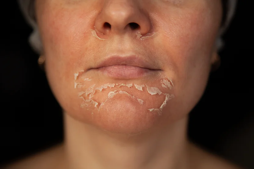
- CHEMICAL PEELS
8.1 Indications
- Epidermal pigmentation
- PIH
- Superficial melasma
- Sun damage
8.2 Safe Peels for Darker Skin
- Mandelic acid
- Lactic acid
- Jessner’s (modified)
- Low-strength TCA (10–15%)
- Azelaic acid peels
8.3 Medium-Depth Peels
Used cautiously in Fitzpatrick IV–VI.
- LASERS & ENERGY-BASED DEVICES
9.1 Principles for Pigmentation
- Start with low fluence
- Avoid aggressive heating
- Protect epidermis
- Consider spot testing
9.2 Q-Switched Lasers
Effective for:
- Lentigines
- Freckles
- PIH (selected cases)
- Dermal pigmentation (Ota, Hori’s)
9.3 Picosecond Lasers
Benefits:
- Faster clearance
- Lower thermal damage
- Better for resistant pigment
9.4 Fractional Non-Ablative Lasers (1550 / 1927 nm)
Useful for:
- Mixed-depth melasma
- Texture + pigment
Avoid overly aggressive settings to prevent rebound.
9.5 IPL (Intense Pulsed Light)
Best for:
- Light skin types (I–III)
- Superficial sun damage
Not recommended for darker skin due to PIH risk.
- REGENERATIVE & ADJUNCTIVE THERAPIES
10.1 Tranexamic Acid (TXA)
Oral TXA
- Reduces plasmin activity
- Controls vascular component in melasma
- Dosage used cautiously under medical supervision
Topical TXA
Useful for melasma and PIH.
Injectable TXA
Emerging option; requires trained professionals.
10.2 Microneedling
Enhances penetration of:
- Vitamin C
- Tranexamic acid
- Glutathione
- Peptides
Useful for PIH and melasma (non-inflammatory approach).
10.3 PRP
Benefits:
- Anti-inflammatory
- Skin repair
- Combination with fractional lasers improves tone
10.4 Exosomes & Cellular Therapies
Modulate:
- Inflammation
- Melanocyte overactivity
- Skin barrier repair
Useful for chronic melasma.
- TREATMENT ALGORITHMS
11.1 Melasma Algorithm
Step 1: Control Triggers
- Sun protection
- Discontinue photosensitizers
Step 2: Topicals (6–12 weeks)
- Hydroquinone
- Retinoid
- Azelaic acid
- Vitamin C
Step 3: Add Procedures
- Gentle chemical peels
- Low-fluence lasers (if stable)
- Microneedling + TXA
- Oral TXA (selected cases)
Step 4: Maintenance
- Non-hydroquinone brighteners
- Strict sun protection
11.2 PIH Algorithm
Primary Goal: Reduce inflammation
- Niacinamide
- Azelaic acid
- Vitamin C
- Gentle AHAs
- Low-strength peels
- Sunscreen
Avoid:
- Ablative lasers
- Aggressive peels
- Heat-based devices
11.3 Lentigines / Sun Damage
- Q-switch or picosecond lasers
- IPL for lighter skin types
- Combination with peels
- SAFETY & COMPLICATION MANAGEMENT
12.1 Post-Inflammatory Hyperpigmentation (PIH)
Most common complication.
Prevention:
- Gentle settings
- Priming skin before treatments
- Sun avoidance
12.2 Hypopigmentation
Due to melanocyte damage.
Avoid aggressive thermal devices.
12.3 Rebound Melasma
Triggered by:
- Heat
- Inflammation
- Over-treatment
Management:
- Return to topical therapy
- Oral TXA in selected patients
12.4 Burns
Usually from lasers or strong peels.
Management:
- Immediate cooling
- Topical steroids (short-term)
- Barrier repair
- PIH prevention
- POST-TREATMENT CARE
- SPF 50+ daily
- Avoid heat exposure
- Avoid exfoliants temporarily
- Gentle skincare only
- Topical antioxidants
- Avoid tanning for minimum 4–6 weeks
Long-term maintenance is essential for chronic conditions.
- KEY LEARNING POINTS
- Hyperpigmentation requires accurate diagnosis before treatment.
- Melasma is chronic and must be managed with long-term plans.
- PIH risk increases significantly in Fitzpatrick IV–VI.
- Topicals are first-line therapy for most pigment conditions.
- Lasers should be used conservatively and selectively.
- Combination regimens outperform monotherapy.
- Sun protection is the cornerstone of prevention and maintenance.
LEARNING OBJECTIVES
After completing this module, learners will be able to:
- Identify and diagnose common types of hair loss.
- Understand the biological mechanisms of PRP, mesotherapy, LLLT, exosomes, and regenerative therapies.
- Develop structured treatment protocols tailored to patient needs.
- Recognize contraindications and high-risk cases.
- Manage complications and set realistic patient expectations.
- INTRODUCTION
Hair loss affects both men and women across various age groups and can significantly impact psychological well-being. Advanced aesthetic medicine provides non-surgical interventions that promote:
- Follicle regeneration
- Improved microcirculation
- Reduction of inflammation
- Activation of dormant follicles
- Hair shaft strengthening
Proper diagnosis and a multimodal approach produce optimal outcomes.
- TYPES OF HAIR LOSS & DIAGNOSTIC WORK-UP
2.1 Androgenetic Alopecia (AGA)
Most common type.
Characterized by:
- Miniaturization of hair follicles
- Shortened anagen phase
- Genetic and hormonal influence (DHT sensitivity)
Typical patterns:
- Male pattern: temples & crown
- Female pattern: diffuse thinning over crown
2.2 Telogen Effluvium (TE)
Trigger-induced shedding.
Causes:
- Stress
- Fever/illness
- Postpartum
- Medications
- Nutrient deficiencies
Reversible when trigger addressed.
2.3 Alopecia Areata
Autoimmune disorder
Patchy hair loss
May require combination therapy with dermatology oversight.
2.4 Scarring Alopecia
Permanent destruction of follicles.
Referral to dermatologist required.
2.5 Diagnostic Evaluation
Clinical Evaluation
- Hair-pull test
- Pattern mapping
- Dermatoscopy (trichoscopy)
Laboratory Investigation
Recommended tests:
- Ferritin
- Vitamin D
- Thyroid profile
- Zinc levels
- Hormone panel (PCOS, androgens if indicated)
Accurate diagnosis guides correct treatment selection.
- PLATELET-RICH PLASMA (PRP)
3.1 Mechanism of Action
PRP contains growth factors including:
- PDGF
- TGF-β
- VEGF
- EGF
These promote:
- Angiogenesis
- Prolonged anagen phase
- Reduced inflammation
- Follicular regeneration
3.2 PRP Preparation
Factors influencing quality:
- Centrifuge type (single vs double spin)
- Platelet concentration
- Use of anticoagulants
Ideal concentration: 4–5x baseline platelet count
3.3 Clinical Protocol
- Sessions: every 3–6 weeks for 3–6 sessions
- Maintenance: every 3–6 months
- Injection depth: 1–3 mm intradermal
- Method: nappage or point-by-point
3.4 Indications
- Androgenetic alopecia (early–moderate)
- Telogen effluvium (as adjunct)
- Hair quality improvement
3.5 Advantages
- Autologous
- Safe
- Minimal downtime
3.6 Limitations
- Requires multiple sessions
- Not effective in advanced miniaturization
- Technique-dependent
- MESOTHERAPY FOR HAIR LOSS
4.1 Mechanism of Action
Mesotherapy delivers microinjections of active ingredients into the dermis.
Formulas commonly include:
- Vitamins (Biotin, B-complex)
- Amino acids
- Peptides
- Hyaluronic acid
- Minerals
- DHT blockers
Mechanisms:
- Improved blood flow
- Nutrient delivery
- Reduced inflammation
- Follicle stimulation
4.2 Treatment Protocol
- Weekly sessions x 4–8
- Maintenance every 1–3 months
- Injection technique: microbolus or nappage
4.3 Indications
- Telogen effluvium
- Early androgenetic alopecia
- Hair shaft strengthening
- LOW-LEVEL LASER THERAPY (LLLT)
5.1 Mechanism
LLLT stimulates mitochondria in follicular cells.
Effects include:
- ATP production
- Increased blood circulation
- Prolonged anagen phase
- Reduced inflammation
5.2 Clinical Use
- 2–3 sessions per week
- Helmet, comb, or panel devices
5.3 Indications
- AGA (male & female)
- Diffuse thinning
- Maintenance therapy
- MICRONEEDLING FOR HAIR RESTORATION
6.1 Mechanism
Microneedling creates controlled micro-injuries:
- Increases growth factor release
- Enhances topical penetration
- Stimulates Wnt/β-catenin pathway
- Supports dermal papilla activation
6.2 Protocol
- Needle depth: 0.6–1.5 mm
- Frequency: every 1–2 weeks
- Often combined with PRP, peptides, or minoxidil
- REGENERATIVE THERAPIES
7.1 Exosomes
Extracellular vesicles containing:
- Growth factors
- Cytokines
- mRNA
- miRNA
Benefits:
- Reduce inflammation
- Activate stem cells
- Restore follicular microenvironment
7.2 Polynucleotides (PN / PDRN)
Mechanism:
- Enhance tissue repair
- Improve blood flow
- Reduce oxidative stress
- Support hair density
7.3 Stem Cell-Based Therapies
Include:
- Adipose-derived stem cells
- Autologous cellular concentrates
Work by regenerating follicular niche.
7.4 Combination Regenerative Protocols
Examples:
- Exosomes + microneedling
- PRP + PN
- PRP + microneedling + LLLT
Combination therapy delivers superior outcomes.
- TOPICAL & SYSTEMIC ADJUNCTS
8.1 Topical Minoxidil
- Vasodilator
- Increases anagen duration
Forms:
- 2%, 5%,
8.2 Finasteride / Dutasteride
5α-reductase inhibitors
Used in male AGA (and selected female cases)
8.3 Multivitamins & Supplements
Beneficial in deficiency-related cases:
- Iron
- Vitamin D
- Zinc
- Biotin
- COMBINATION APPROACHES
9.1 Examples of Integrated Treatment Plans
Male AGA (Early Stage)
- PRP every 4 weeks x 3
- Oral finasteride
- Minoxidil topical
- LLLT 2–3x weekly
Female Pattern Hair Loss
- PRP x 3
- Mesotherapy with peptides
- Topical minoxidil
- Address hormonal imbalances
Telogen Effluvium
- Mesotherapy
- Nutritional correction
- Stress management
- Maintained with PRP or LLLT
Advanced AGA
- Combined PRP + exosomes
- Microneedling every 2 weeks
- Consideration of transplantation
- SAFETY, RISKS & COMPLICATION MANAGEMENT
10.1 PRP
- Bruising
- Mild pain
- Swelling
- Infection (rare with sterile technique)
10.2 Mesotherapy
- Allergic reactions (depending on formula)
- Swelling
- Tenderness
- Rare nodules
10.3 LLLT
Extremely safe
Occasional headaches or scalp warmth
10.4 Microneedling
- Pinpoint bleeding
- Erythema
- PIH in dark skin if overly aggressive
10.5 Exosomes / PN
- Mild redness
- Temporary sensitivity
- POST-TREATMENT CARE
General recommendations across treatments:
- Avoid sweating for 24 hours
- Avoid hair dye for 48–72 hours
- No heavy exercise for 24 hours
- Use gentle shampoo
- No heat exposure
- Avoid anti-inflammatory medications for 24 hours after PRP
Follow-up every 4–6 weeks is essential for tracking progress and adjusting the plan.
- KEY LEARNING POINTS
- Hair loss requires proper diagnosis before treatment begins.
- PRP is a cornerstone treatment with strong regenerative effects.
- Mesotherapy improves scalp nutrition and complements other therapies.
- LLLT enhances cellular metabolism and supports long-term maintenance.
- Exosomes and PN offer advanced biologic rejuvenation.
- Combination therapy provides superior results over monotherapy.
- Patient education and compliance determine long-term success.
Related Posts
Chemical Peels in Aesthetic Medicine
Posted on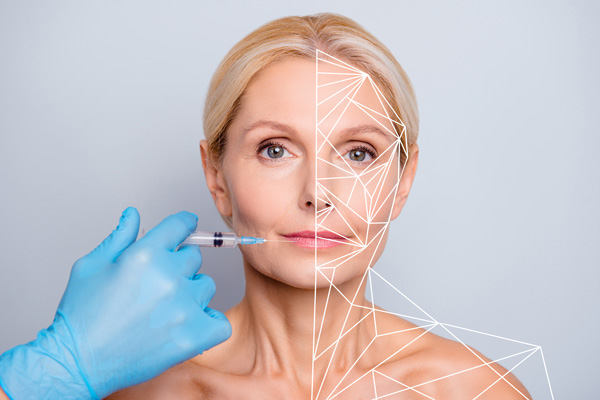
Complications in Aesthetic Medicine
Posted on