ABSTRACT
Dermal fillers play a central role in facial rejuvenation and harmonization through volume restoration, contour enhancement, and structural support. Their effectiveness depends heavily on precise anatomical knowledge, appropriate product selection, safe injection techniques, and proficient management of complications. This educational module provides an in-depth foundation covering filler classifications, rheology, facial anatomy, treatment planning, injection protocols, complication management, and best practices for safe and natural results.
LEARNING OBJECTIVES
After completing this module, learners will be able to:
- Understand the classification and rheological properties of dermal fillers.
- Recognize essential facial anatomy, fat compartments, and vascular structures.
- Apply appropriate indications and treatment planning for facial enhancement.
- Perform safe injection techniques for each anatomical region.
- Identify early warning signs of complications and manage them effectively.
- Develop individualized patient-centered treatment strategies.
- INTRODUCTION
Dermal fillers are injectable materials designed to restore volume, contour, hydration, and structural integrity to the face. They are widely used for:
- Midface volumization
- Jawline contouring
- Chin projection
- Lip enhancement
- Tear trough correction
- Non-surgical rhinoplasty
- Fine-line and superficial corrections
Hyaluronic acid (HA) fillers are the most commonly used due to their reversibility and safety profile. Other fillers include calcium hydroxyapatite, poly-L-lactic acid (PLLA), and PMMA, each with distinct indications, longevity, and handling characteristics.
Successful filler treatment requires deep understanding of facial anatomy, aging patterns, rheology, and safe injection techniques.
- FILLER MATERIALS & RHEOLOGY
2.1 Hyaluronic Acid (HA) Fillers
- Most widely used
- Hydrophilic, reversible with hyaluronidase
- Different cross-linking levels create diverse gel consistencies
Used for:
• Lips • Tear troughs • Cheeks • Jawline • Nasolabial folds • Lines & wrinkles
2.2 Calcium Hydroxyapatite (CaHA)
- Semi-permanent
- Biostimulatory
- Strong lifting capacity
Used for:
• Jawline • Midface • Lower face tightening • Collagen stimulation
2.3 Poly-L-Lactic Acid (PLLA)
- Collagen stimulator
- Requires multiple sessions
Used for:
• Global facial volume loss • Skin tightening • Long-term collagen remodeling
2.4 PMMA (Polymethyl Methacrylate)
- Permanent
- Requires advanced experience due to higher complication risk
Used for:
• Deep volume restoration • Structural support
- FACIAL ANATOMY FOR DERMAL FILLER INJECTIONS
Mastery of anatomy is the most critical component of safe filler practice.
3.1 Skin & Soft Tissue Layers
- Epidermis
- Dermis
- Superficial fat compartments
- SMAS
- Deep fat compartments
- Periosteum
Aging affects each layer differently, resulting in volume deflation, ligament laxity, and bone remodeling.
3.2 Facial Fat Compartments
Facial fat is divided into superficial and deep compartments with distinct functions:
Superficial Compartments
- Mobile
- Influence facial expression
- Ideal for fine-line corrections or soft contouring
Deep Compartments
- Provide foundational support
- Best for structural lifting and projection
Common deep compartments treated with fillers:
- Deep medial cheek fat
- Lateral cheek fat
- Pre-maxillary space
- Chin fat compartments
- Pre-jowl sulcus
3.3 Vascular Anatomy: Critical Danger Zones
Understanding vascular anatomy is essential to avoid severe complications.
High-risk zones include:
- Glabella (supraorbital & supratrochlear vessels)
- Nose (dorsal and lateral nasal arteries)
- Nasolabial fold region
- Tear trough & infraorbital area
- Temple (superficial temporal artery)
- Lips (superior & inferior labial arteries)
Knowledge of vessel depth, course, and variations reduces risk of intravascular injection.
- AESTHETIC INDICATIONS
4.1 Upper Face
- Temporal hollowing
- Eyebrow support
- Forehead contouring (advanced)
4.2 Midface
- Cheek augmentation
- Malar projection
- Tear trough correction
- Nasolabial fold softening
- Pre-maxillary augmentation
4.3 Lower Face
- Lips (shape, volume, hydration)
- Marionette lines
- Chin definition and projection
- Jawline contouring
- Pre-jowl sulcus correction
4.4 Nose (Non-Surgical Rhinoplasty)
Specialized treatment requiring high skill due to vascular risk.
4.5 Skin Quality Improvement
- HA skin boosters
- Fine-line intradermal fillers
- TREATMENT PLANNING & PATIENT SELECTION
5.1 Patient Assessment
Include:
- Medical history
- Previous filler treatments
- Allergies
- Skin quality
- Facial asymmetry
- Dynamic vs static wrinkles
- Aging stage (per decade)
5.2 Aesthetic Goals
Define patient’s goals clearly:
- Volume
- Lifting
- Contouring
- Hydration
- Symmetry
5.3 Contraindications
- Active infection or inflammation
- Pregnancy/breastfeeding
- Autoimmune syndromes (relative)
- Unrealistic expectations
- Recent dental procedures (relative)
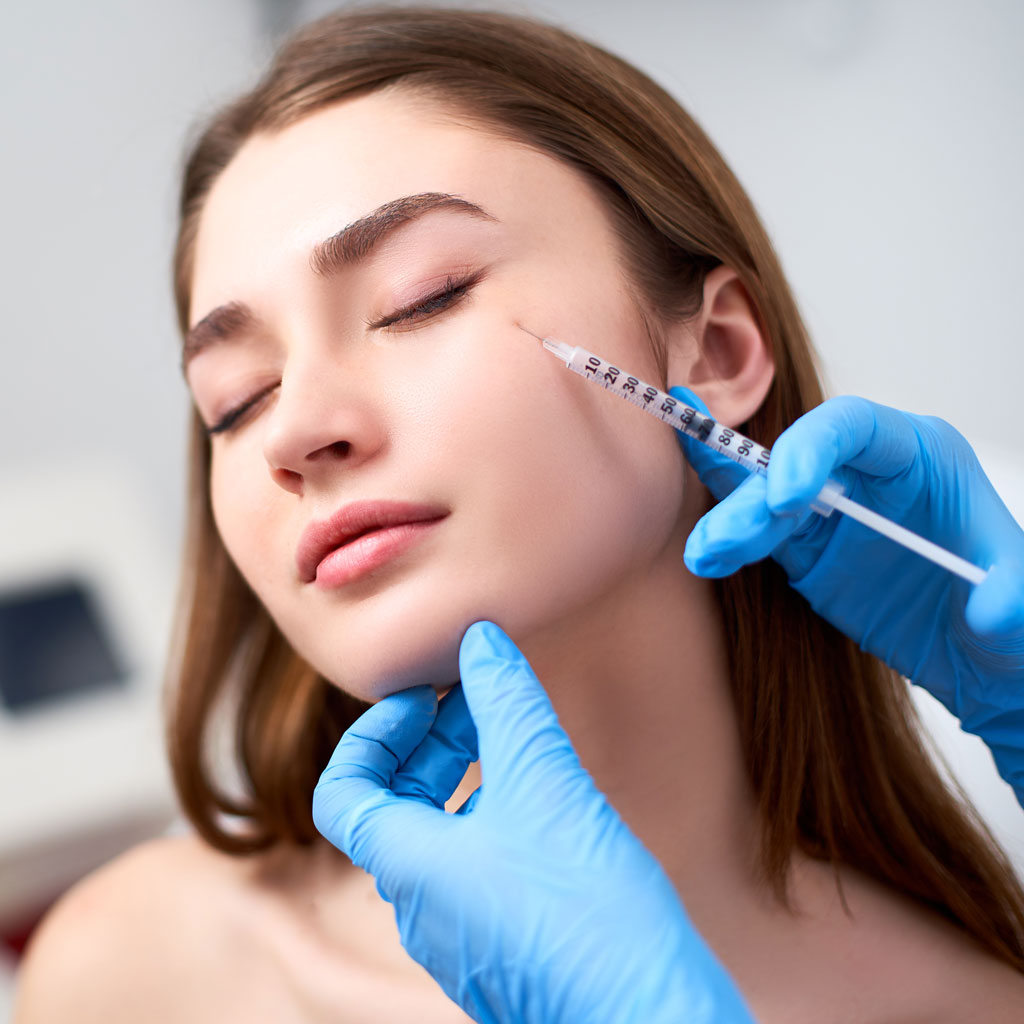
- INJECTION TECHNIQUES & SAFE PRACTICE
6.1 Needle vs Cannula
Needle
- Precise placement
- Best for structural areas
Cannula
- Lower risk of vascular penetration
- Ideal for sensitive/high-risk areas
6.2 Injection Depth per Region
Cheeks
- Deep bolus on bone
- Purpose: lifting and contouring
Tear Trough
- Deep, supraperiosteal or deep-fat plane
- Low G’ fillers recommended
Lips
- Submucosal or vermillion border
- Microdroplet or linear threading
Jawline
- Deep supraperiosteal for structure
- Cannula for smooth contouring
Chin
- Deep bolus for projection
- Horizontal/vertical augmentation
Temple
- Deep (beneath deep temporal fascia)
- Cannula preferred for safety
6.3 General Technical Principles
- Inject slowly with low pressure
- Aspirate where necessary
- Use minimal effective volume
- Constantly monitor skin color & patient feedback
- Avoid overcorrection
- Stop immediately if pain, blanching, or livedo occurs
- COMPLICATIONS & MANAGEMENT
Complications can range from mild to severe. Early recognition saves tissue and prevents long-term issues.
7.1 Expected Minor Events
- Bruising
- Swelling
- Tenderness
- Asymmetry
- Irregularities
Typically resolve within days.
7.2 Immediate Complications
Intravascular Injection (Vascular Occlusion)
Signs:
- Severe pain
- Blanching
- Mottled skin (livedo reticularis)
- Vision changes (in nose/glabella injections)
Management Principles:
- Stop injection immediately
- Inject high-dose hyaluronidase in affected area
- Warm compresses
- Massage
- Aspirin (per local protocol)
- Monitor continuously
Vision changes require urgent ophthalmology referral.
Nodules & Lumps
Types:
- Inflammatory
- Non-inflammatory
Management:
- Massage
- Warm compress
- Hyaluronidase if HA-related
- Antibiotics if infection suspected
Tyndall Effect
Blue hue due to superficial placement.
Management: hyaluronidase.
7.3 Delayed Complications
Biofilm Formation
- Chronic low-grade infection
- Triggered by trauma, illness, or additional procedures
Management:
- Oral antibiotics
- Possible hyaluronidase
- Complete filler dissolution if recurrent
Delayed Hypersensitivity Reactions
Management:
- Antihistamines
- Corticosteroids
- Consider dissolving filler
- POST-TREATMENT CARE
- Avoid heavy exercise for 24 hours
- Avoid touching or massaging the area
- Avoid makeup for 12 hours (unless intradermal boosters)
- No sauna, steam, or facials for one week
- Reassess at 2 weeks
- Encourage hydration and good skincare
Follow-up is essential to evaluate symmetry and refine results.
- KEY LEARNING POINTS
- Dermal fillers require advanced anatomical knowledge and controlled technique.
- Understanding rheology helps match filler properties to each indication.
- Safe injection depth varies by region and must be respected.
- Early recognition of vascular occlusion saves tissue and prevents scarring.
- Slow injection, small aliquots, and patient feedback reduce complication risk.
- Long-term patient satisfaction depends on natural, individualized treatment planning.

mbakr